Project:
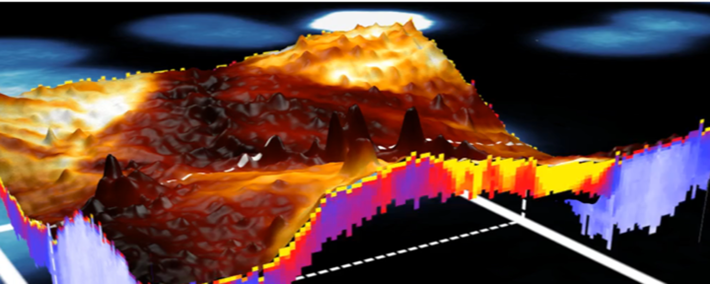
In the directed pharmacotherapy nanoparticle drugs accumulate inside tumour cells – but the occurring processes are not fully explored yet. Magnetic force microscopy is used to subsurface-detect such core-shell structured nanoparticles inside cancer cells. Using multimodal excitation, different physical observables are accessible at the same time, such as topography, magnetic field or Young’s modulus. A frequency modulated control enables high imaging speed and at the same time high resolution imaging. Main goal of this project is to establish the connection between the favoured sites of permeation of the nanoparticles and the mechanical properties of the tumour cells, by providing new, enhanced AFM techniques.
Publications
- A Anahid, FD Hastert, LO Heim, C Dietz (2020)
Reliability of cancer cell elasticity in force microscopy
Appl. Phys. Lett. 116, 083701
DOI: https://aip.scitation.org/doi/10.1063/1.5143432
[Artikel]
- A Anahid, FD Hastert, C Dietz (2020)
Carcinomas with Occult Metastasis Potential: Diagnosis/Prognosis Accuracy Improvement by Means of Force Spectroscopy
Advanced Bio Systems, Volume 4, Issue 7
DOI: https://onlinelibrary.wiley.com/doi/full/10.1002/adbi.202000042
[Artikel]
- Amiri, Anahid and Hastert, Florian D. and Stühn, Lukas and Dietz, Christian (2019):
Structural Analysis of Healthy and Cancerous Epithelial Breast Type Cells by Nanomechanical Spectroscopy Allows to Obtain Peculiarities of Skeleton and Junctions.
In: Nanoscale Advances, ISSN 2516-0230,
DOI: 10.1039/C9NA00021F,
[Artikel] - Stühn, Lukas and Auernhammer, Julia and Dietz, Christian (2019):
pH-depended protein shell dis- and reassembly of ferritin nanoparticles revealed by atomic force microscopy.
9, In: Scientific Reports, 2019 (1), ISSN 2045-2322,
DOI: 10.1038/s41598-019-53943-3,
[Artikel] - Stühn, Lukas and Fritschen, Anna and Choy, Joseph and Dehnert, Martin and Dietz, Christian (2019):
Nanomechanical sub-surface mapping of living biological cells by force microscopy.
In: Nanoscale, ISSN 2040-3364,
DOI: 10.1039/C9NR03497H,
[Artikel]


